X-ray imaging
Summary
We develop and use novel methods for X-ray imaging with the highest possible spatial and temporal resolution.
To this end, we use a wide range of X-ray imaging methods at different synchrotrons, including MAX IV in Lund, X-ray free-electron lasers, including European XFEL, as well as lab source systems. We collaborate with many other researchers, in particular at Lund University, MAX IV, and internationally.
Nanotomography at MAX IV

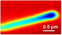
We have established in-line holography at MAX IV, a technique capable of studying nature in 2D and 3D from the microscale down the nanoscale. In-line holography exploits the unique capabilities of MAX IV and the exceptionally small focus of NanoMAX beamline to bridge the capabilities of optical and electron microscopes. This high spatial and temporal resolution enables in-situ and operando imaging experiments at NanoMAX. The video above shows the 3D reconstruction of a cellulose fiber at a resolution of around 100 nm (10 million times smaller than a meter).
Contact
Pablo Villanueva Perez' contact information in Lund University's Research Portal
Phase contrast tomography
We have built a phase contrast X-ray tomograph based on a microfocus Cu source. Traditional X-ray imaging is based on absorption contrast, which has poor contrast for small and weakly absorbing samples. Much better contrast can be achieved using phase contrast. The system is based on a microfocus X-ray source with a Cu target, a high-resolution detector and a high-precision sample stage. It's capable of X-ray absorption contrast and phase contrast imaging at a spatial resolution of about 1 micrometer in 2D and 3D. The image above shows part of a blueberry seed.
Contact
Jesper Wallentin's contact information in Lund University's Research Portal
X-ray imaging with artificial intelligence

Artificial intelligence (AI) has become a key ingredient of computer vision. In our group, we exploit and develop AI algorithms to address specific X-ray imaging problems in 2D, 3D, and 4D (3D+time). One of such problems is the radiation damage that limits the achievable resolution and can be harmful. For that purpose, we have developed AI algorithms that reduce the radiation damage on the sample while keeping the image quality. Furthermore, these algorithms are extremely fast, enabling real-time data analysis. The video above shows the AI reconstructed movie of the coalescence of a metallic foam filmed at MHz rate with an X-ray microscope.
Contact
Pablo Villanueva Perez' contact information in Lund University's Research Portal
Coherent X-ray imaging of nanostructures
The spatial resolution in traditional X-ray imaging is limited by the quality of the optics. In coherent X-ray imaging, the diffracted signal from the sample is analyzed using advanced algorithms, which makes it possible to improve the spatial resolution. We develop methods for coherent X-ray imaging, both in transmission and Bragg geometry, and combine this with electrical measurements.
Contact
Jesper Wallentin's contact information in Lund University's Research Portal
Interested in working with us?
Available BSc/Msc projects
We welcome candidates for BSc and MSc thesis.
PhD and PostDoc positions
When available published on Lund University's website. Direct contact is welcome.
Project leaders
Links will open in Lund University's Research Portal
Postdocs / PhD students
- Hanna Dierks
- Susanna Hammarberg
- Yuhe Zhang